

#1 Intraoral view of pre surgery

#2 Lateral view of teeth absence in posterior maxilla

#3 Intraoral occlusal view
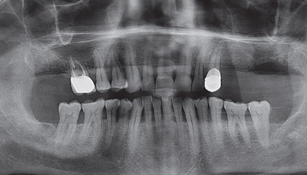
#4 Panoramic view of pre surgery

#5 Opened bone window for sinus lift

#6 Preparation of implant site

#7 Placed 2 Mode dental implant

#8 I-PRF was applied to the bone greft

#9 Placed Mode dental implants

#10 A-PRF and I-PRF were applied to sinuslift area

#11 Panoramic view of post surgery

#12 Periapical radiograph view of 2 years followed up
60 years old male patient was referred to our oral and maxillofacial surgery department about complaining of dental infection in the left posterior region of the maxilla. He was a diabetic patient and his HbA1c level was 11.2 (mmol/mol). Patient was consulted to endocrinology department after the endocrinology treatment HbA1c level were increased and infected teeth were extracted under the prophylaxis antibiotics protocol.After 3 months the patient’s HbA1c level was decreased to the 7.1 (mmol/mol) and the implant treatment was decided. We planned to place implant with sinus lift at the same surgery. A-PRF and I-PRF were applied to sinuslift according to the Choukroun protocol. 2 MODE implants (3.7×11.5, 4.7×8 mm) were placed to the maxillar left molar area. 6 month later implant crown was placed by prosthodontist. After 2 years followed up, a periapical radiograph was taken to check bone level of implant. According to the clinical and radiographic examination results, there was no bone resorption around MODE dental implants.